
Page 46 - REBRAME - Revista Brasileira de Medicina de Emergência
P. 46
Fortes H.M.S. et al.
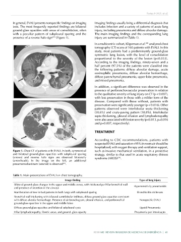
In general, EVALI presents nonspecific findings on imaging Imaging findings usually bring a differential diagnosis that
tests. The most frequently reported findings are bilateral includes infection and a variety of patterns of acute lung
ground-glass opacities with areas of consolidation, often injury, including pneumonia and diffuse alveolar damage.
with a peculiar pattern of subpleural sparing and the The main imaging findings and the corresponding lung
presence of a reverse halo sign [25] (Figure 1). injury are summarized in (Table 1).
In a multicentric cohort, Kligerman et al. [28] analyzed chest
tomography (CT) scans of 160 patients with EVALI. In this
study, most patients had a predominantly ground-glass
symmetric lung lesion, with the level of consolidation
proportional to the severity of the lesion (p<0.033).
According to the imaging findings, ninety-seven and a
half percent (97.5%) of the patients were classified into
the following patterns: diffuse alveolar damage, acute
eosinophilic pneumonia, diffuse alveolar hemorrhage,
diffuse parenchymal pneumonia, upper lobe pneumonia,
and mixed pneumonia.
In addition, a significant difference was observed in the
presence of peribronchovascular preservation in relation
to the qualitative severity of lung injury on CT (p < 0.007),
with less preservation in those with a milder form of the
disease. Compared with those without, patients with
preservation were significantly younger (p < 0.016). Other
patterns observed were interlobular septa thickening
(50.6%) and crazy-paving pattern (18.8%). Interlobular
septa thickening, pleural effusion and lymphadenopathy
were also associated with lesion severity (p<0.013, p=0.016
and p=0.007, respectively).
TREATMENT
According to CDC recommendations, patients with
suspected EVALI and saturation <95% in room air should be
hospitalized, with oxygen therapy and ventilation support,
Figure 1. Chest CT of patients with EVALI. In both, symmetrical such as invasive mechanical ventilation, in a protective
and bilateral ground-glass opacities with subpleural sparing strategy, similar to that used in acute respiratory distress
(arrows) and reverse halo signs are observed bilaterally syndrome (ARDS) [29] .
(arrowheads). In the image on the left, an additional
pneumomediastinum (asterisk) is observed [27] .
Table 1. Main presentations of EVALI on chest tomography.
Image finding Type of lung injury
Bilateral ground-glass changes in the upper and middle zones, with thickened perihilar bronchial wall Hypersensitivity pneumonitis
and presence of secretion in the airways
Manifestation of tree-in-bud patterns in both lungs with subpleural sparing Bronchiolitis obliterans
Bronchial wall thickening with bilateral centrilobular infiltrate, diffuse ground-glass opacities consistent
with diffuse alveolar hemorrhage. Presence of air bronchogram, pleural effusion, and peribronchial Nonspecific EVALI
ground-glass opacities in the upper and middle lobes
Diffuse ground-glass opacities and bilateral subpleural cysts Lipoid Pneumonia
Hilar lymphadenopathy, fibrotic areas, and ground- glass opacity Pneumonia por intoxicação
REBRAME | REVISTA BRASILEIRA DE MEDICINA DE EMERGÊNCIA | 41